Brain Imaging after an Injury

Summary
Emergency medicine physicians make decisions about when and what type of imaging studies to order based on clinical practice guidelines. Learn about the kinds of imaging techniques used assess brain injuries.
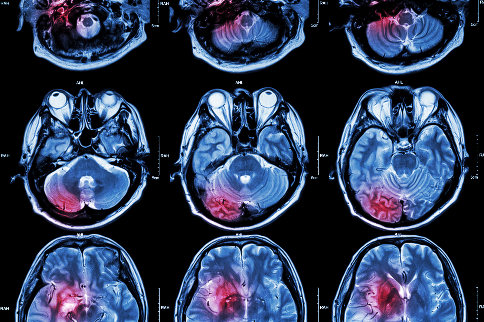
What kind of imaging techniques assess brain injuries? In the old days, the X-ray was the only way to get a look inside someone without surgery. Now, thanks to advances in technology, doctors have an array of imaging methods to see what’s going on. The current primary imaging modalities for assessing brain injuries are Computed Tomography scans (CAT or CT scans) and Magnetic Resonance Imaging scans (MRI scans).
Not everyone who has sustained a blow to the head will, or should, undergo an imaging study. Emergency medicine physicians make decisions about when to order imaging studies based on clinical practice guidelines. (See my pocket guide for more about this.)
When Imaging Is Used After A Suspected Brain Injury
Doctors typically order CT scans after possible damage to the structure of the skull or brain, when they suspect bleeding in the brain, in order to decide if surgery is necessary. MRI scans are not often ordered in the early, acute period even with more severe injuries. An MRI scan may be ordered later, if symptoms persist after a concussion.
Concussions or mild brain injuries don’t usually show evidence of injury on standard MRI and CT scans, the most commonly used types of imaging for brain injuries. Physical and cognitive examinations are more reliable ways to diagnose (and plan treatment for) concussions and other brain injuries.
Common Brain Imaging Techniques
The following list includes the common types of imaging used to determine whether the head or brain has sustained an injury.
X-rays (radiographs) for TBI
Radiographs are images created using X-rays, and this remains the most common type of imaging technology in medicine. X-rays are not typically ordered if a CT scan is performed. X-rays reveal bone structures and are ordered to further evaluate fractures of the face, head, and skull.
X-rays (electromagnetic radiation) are emitted towards the affected body part. Some X-rays are absorbed and others pass through to the film or detector on the other side of the body, creating a 2-D image.
CT Scans (CAT Scans) for Brain Injury
CT scans also use X-ray technology. However, unlike radiographs, CT images offer a better understanding of the whole picture, because tomographic (that is, cross-sectional) images show “slices” from portions of the brain or other parts of the body. While X-rays for a radiograph come from a single direction, X-rays for CT scans use a rotating X-ray generator that circles the head or body to gather multiple images.
CT scans are the standard imaging choice when a brain injury is suspected because the technology is readily available in hospitals and outpatient centers. CT scans can show skull and facial fractures as well as swelling, bleeding, or foreign objects.
MRI Scans for Brain Injury
MRI scans use magnetic fields and radio waves to create images that, like CT scans, are also a collection of “slices” of the brain. Compared to CT scans, MRIs take longer and are often less comfortable for the patient, but they can show more detailed and different information than a CT scan. For instance, MRIs can show changes in the brain such as bruising and scarring that are not detectable with CT imaging, and they can also show even smaller areas of bleeding in the brain such as microhemorrhages seen in diffuse axonal injury (DAI).
How does it work? The MRI scanner produces a strong magnetic field to affect hydrogen nuclei in the body and then uses radio waves (another form of electromagnetic radiation) to get information from those nuclei. It’s the radio waves that produce the images we see in the final MRI scans. (For a more in-depth explanation and for the different kinds of MRI scanning techniques, go here.)
fMRI Scans for Brain Injury
Functional MRI (fMRIs) record activity in the brain during mental tasks. They are primarily used for research. fMRIs use the same magnetic resonance imaging as MRIs, but rather than using hydrogen nuclei, fMRI takes advantage of the fact that oxygen-rich hemoglobin in the blood acts differently in a magnetic field than oxygen-poor hemoglobin. That enables the fMRI imaging to look at real-time blood flow in the brain.
PET Scans for TBI
Positron Emission Tomography (PET) is used in brain injury research. PET scans are similar to fMRI scans – both are “dynamic imaging modalities” that look at brain activity, rather than the structure of the brain. To produce PET images, radioactive chemicals that emit positrons are injected into the bloodstream and the detector creates images that reflect brain activity. The assumption is that blood flow or oxygen use, for example, correlates with activity.
Learn More About Brain Injuries
I discuss what happens to the brain after trauma in my book, Shaken Brain: The Science, Care, and Treatment of Concussion (see especially Chapters 1 and 4).
My blog on chronic traumatic encephalopathy (CTE) addresses a condition caused by repetitive concussions and subconcussive “hits” to the brain that occur in football and other collision sports.
You might also be interested in my podcasts about concussion and other traumatic brain injuries, with Ronit Molko.
You Might Also Like
The Prevalence of Brain Injuries in Sports and the Military
In the second part of this series, Dr. Sandel continues with further discussion of concussion management. She then describes blast injuries that occur in the military. Who provides treatment for concussions and what kind of care is best?, What are the risks of a long-term problem after a concussion or…
The “Invisible Injury”: Concussions and other TBI’s
In the first part of this series, Dr. Sandel discusses mild brain injuries, especially sports-related concussions. What happens to the brain during a concussion and what are the symptoms? What is second impact syndrome? And are children more or less vulnerable than adults?
Repetitive Brain Trauma and Chronic Traumatic Encephalopathy (CTE)
There’s a link between chronic traumatic encephalopathy (CTE) and repetitive brain injuries that occur in boxing and American football. This is a progressive neurodegenerative disorder that can lead to severely-disabling neurologic and psychiatric disorders. Learn about the science, diagnostic criteria for traumatic encephalopathy syndrome (TES), and possible treatment approaches.
Keep up to date
Get updates on the latest in concussion, brain health, and science-related tools from Dr. Elizabeth Sandel, M.D.
By clicking SIGN UP, you agree to receive emails from Dr. Sandel and agree to our terms of use and privacy policy.
Get the book!


